Ophthalmology
Acuity, pressure and the retina — findings placed on the eye itself.
Four kinds of visit — routine and refraction, the red or painful eye, glaucoma and diabetic screening. Findings are placed on real photographs of the outer eye, the anterior segment and the fundus, right and left.
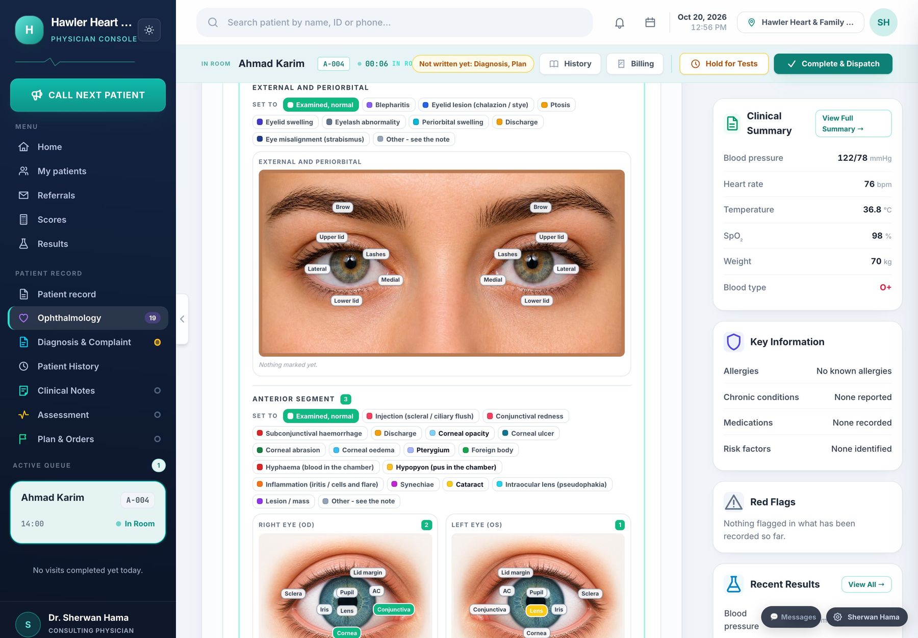
Findings on the eye: lids, conjunctiva, cornea and lens on photographs, both eyes.
Visual acuity, refraction, pressure, pupils, the anterior segment and the fundus, with diabetic and glaucoma pathways.
Acuity and refraction
Unaided, corrected and pinhole for each eye; sphere, cylinder, axis and spherical equivalent.
Pressure and pupils
Intraocular pressure and method, pachymetry, pupil reactions, RAPD, motility and squint.
Anterior segment and fundus
Cataract, ulcer, iritis, haemorrhage, exudates, new vessels, disc cupping — marked on the structure.
Diabetic retinopathy
Grading for each eye, maculopathy, laser and injections, and when screening is next due.
The details that make the difference.
Everything on this page is in the software today. Nothing here is a promise for later.
- ✓Fluorescein, Van Herick, tear break-up time, Schirmer and gonioscopy
- ✓Cup-to-disc ratio for each eye, for glaucoma follow-up
- ✓The marked eye prints into the visit report
One record, shared by every room.
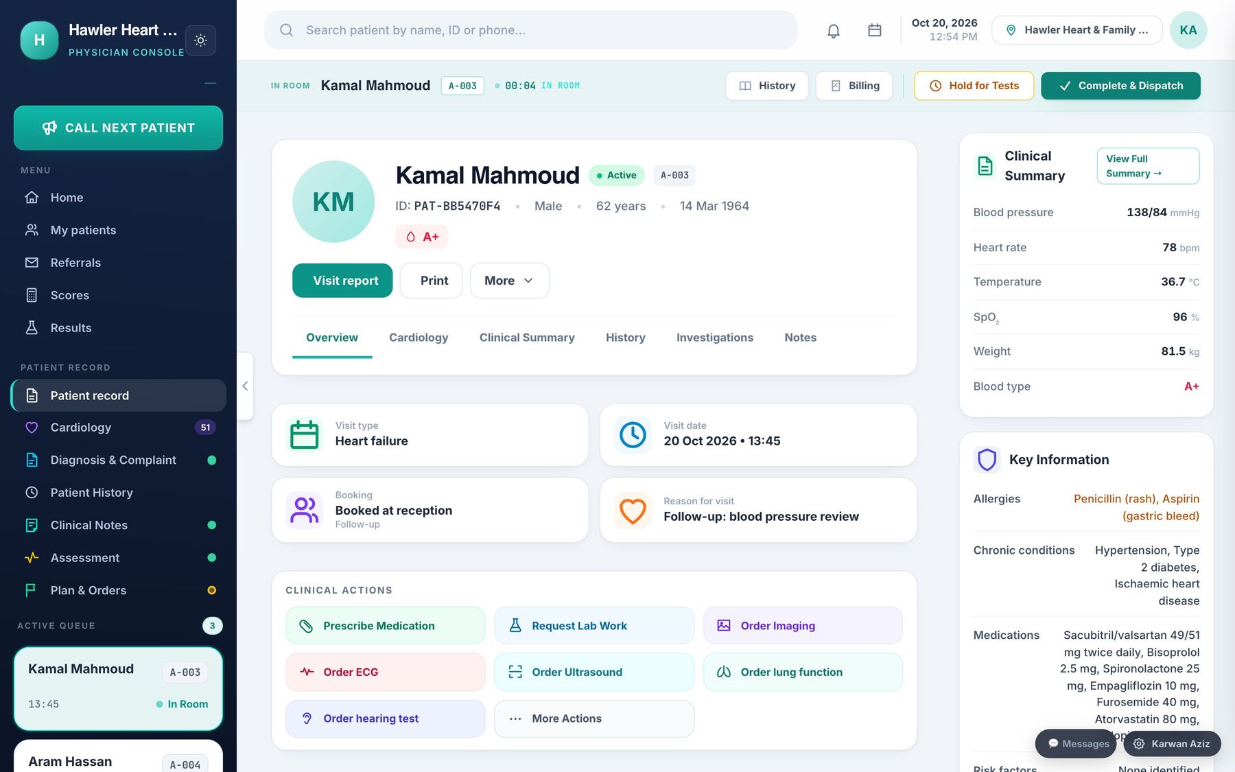
The doctor’s console
One screen for the consultation: notes, specialty chart, orders, prescriptions with allergy checks, and the visit report.
Read more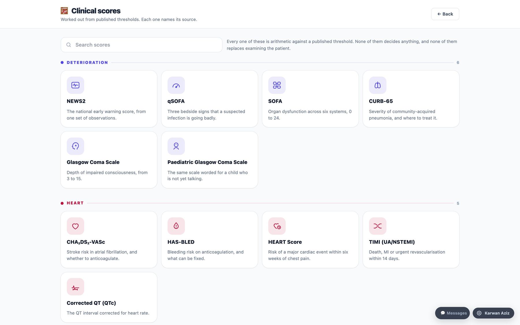
All 22 specialties
From general practice to oncology — 22 specialty charts and 31 clinical scores, each named with its source.
Read moreSafety, privacy & backups
Allergy and critical-result safety, roles, the audit trail, clinic separation, backups and three languages.
Read more